Scientific background Charged Multiscan.
How it works
The Multiscan is based on functional analysis and physiological data – not to be confused with bioenergetic scans or bioresonance techniques. It is a computer-controlled system that visualizes collected data using scientifically validated technologies. These visualizations consist of images, diagrams, and customized graphs that provide valuable insights into how your body works and your overall health. The results serve as a baseline for monitoring and as a reference point for tracking your progress over time.
Below you will find an explanation and scientific background of the technologies used by the Multiscan:
Heart rate variability (HRV)
Digital pulse wave analysis (DVPA)
Galvanic skin response (GSR/EDA)
Bioelectrical impedance analysis (BIA)
Heart Rate Variability (HRV)
HRV measures the fluctuations in the time interval between successive heartbeats, expressed in milliseconds (ms). It is an important indicator of the flexibility and adaptability of the autonomic nervous system, and reflects the balance between:
The parasympathetic nervous system (rest and digest), which slows down the heart rate and increases variability (higher HRV).
The sympathetic nervous system (fight-or-flight), which accelerates the heart rate and reduces variation (lower HRV).
HRV is widely considered to be the most effective method for assessing the balance of the autonomic nervous system, due to its direct influence on heart activity and broad scientific basis. However, HRV is highly individual, making comparisons between individuals difficult. Regular HRV measurements help to provide insight into personal trends and developments over time.
Scientific references:
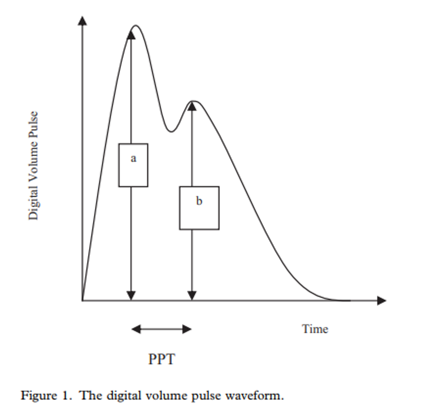
Digital Volume Pulse Analysis (DVPA)
Photoplethysmography (PPG) is a non-invasive technique that measures changes in blood volume using optical sensors.
This method is often used to determine heart rate and blood oxygen levels and provides valuable insights into cardiovascular health.
The shape of the pulse waves reveals information about arterial stiffness, vascular resistance, and overall cardiovascular function.
The digital volume pulse (DVP), similar to the pressure pulse, responds to changes in arterial tension and aging. By analyzing this pulse, we can assess the properties of the blood vessels.
The arterial pulse wave consists of two main components: a forward wave and a reflected wave. The speed of the reflected wave is an important indicator—stiffer blood vessels cause faster reflection, which affects systolic blood pressure.
Scientific references:
Galvanic Skin Response (GSR) / Electrodermal Activity (EDA)
GSR measures the electrical conductivity of the skin in response to external stimuli.
It is a physiological indicator controlled by the autonomic nervous system, particularly the sympathetic nervous system.
When someone is emotionally aroused, anxious, or stressed, the activity of the sympathetic nervous system increases. This leads to higher sweat production and changes in skin conductivity. The galvanic skin response (GSR) is a well-documented and scientifically validated measure of physiological arousal. The term electrodermal activity (EDA) is often used as a synonym.
Scientific references:
Bioelectrical Impedance Analysis (BIA)
During a multisegmental BIA measurement, weak electrical currents are passed through the body. Different tissues—such as fat, muscle, bone, and water—each offer a different degree of resistance. By measuring these resistances, the BIA can estimate body composition.
The results of the BIA provide insight into, among other things:
Fat percentage
Muscle mass
Total body water
Other important health indicators
The BIA is widely used in healthcare and the fitness world to assess and monitor a person's health and fitness over time.
Scientific references: